Q: I am home for the summer and have been focusing on getting my horse fit again. Even though I have been taking it slow and steady, he doesn’t seem to have the same stamina this year. Is this something I should be concerned about? If so, what can I do to address it?
A:
A horse experiencing a change in his tolerance for exercise may have an underlying health issue. A thorough physical examination by your regular veterinarian can point you in the direction of getting the therapy your horse needs.
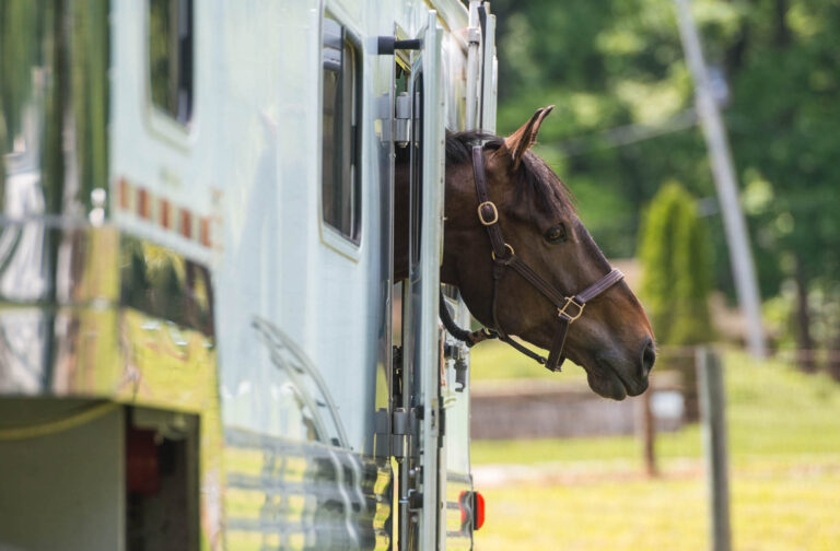
When faced with a concern about exercise intolerance, the first body system we focus on is the respiratory tract. Respiratory disease can contribute to exercise intolerance due to an insufficient ability to supply oxygen to support athletic activity. We generally categorize these diseases as originating either in the upper or lower airways.
In the upper airways, this problem is largely due to an anatomical change in the horse’s larynx, which hinders airflow. A recognizable disease in this category is laryngeal hemiplegia or “roaring.” Alternatively, the problem can originate in the lower airways if there is inflammation spread throughout the lung tissue that impairs the ability of oxygen to get into the blood that then circulates through the body. While upper-airway obstructive diseases are typically associated with an audible noise, the early signs of diffuse lower-airway inflammatory disease are more subtle, such as flared nostrils and a higher than normal respiratory rate even at rest.
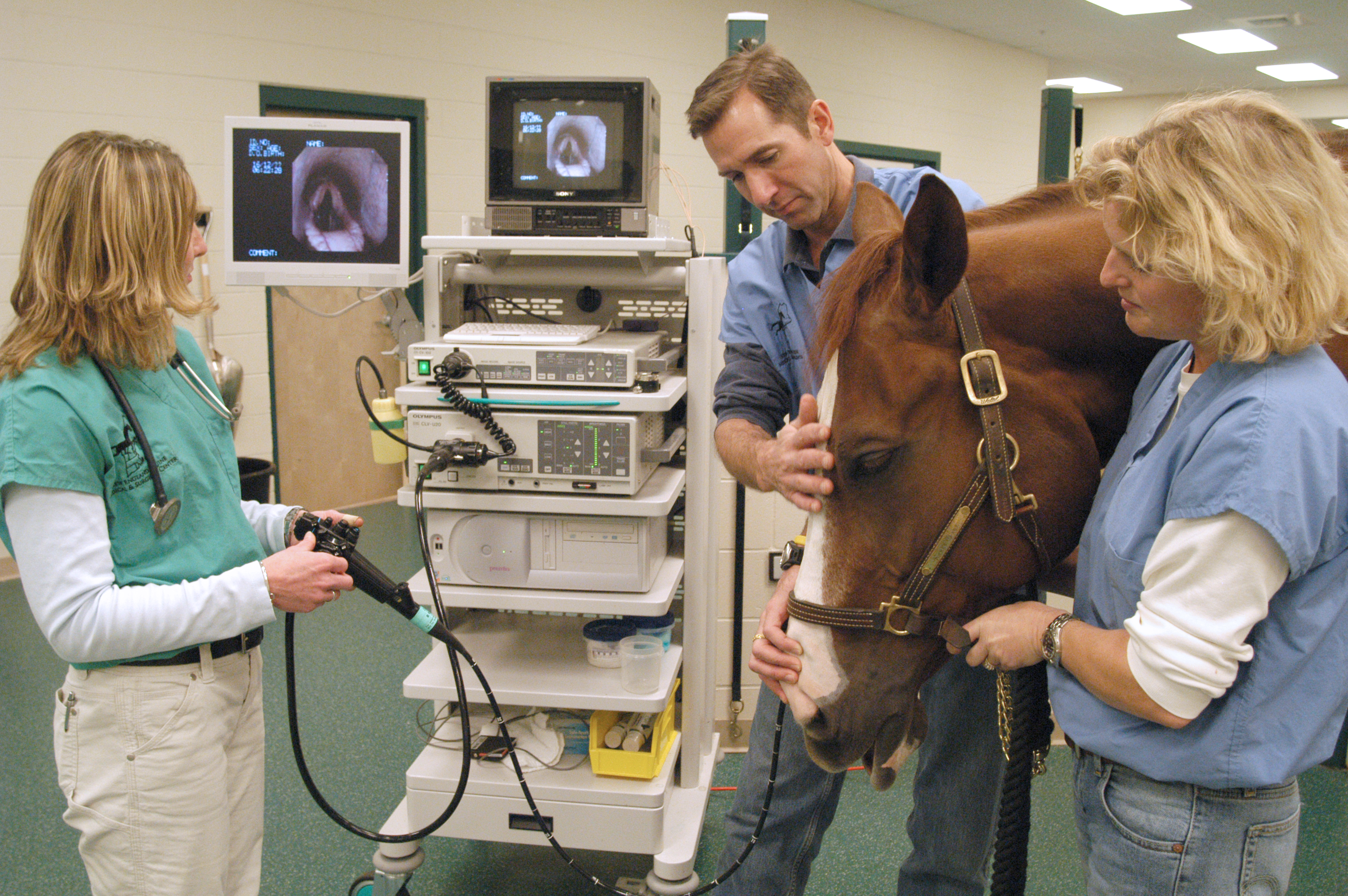
There are two basic diagnostic steps to assess the respiratory tract: an upper- airway endoscopy and a rebreathing exam. Both procedures are relatively well tolerated by horses and can be done in the barn. In an endoscopic exam, a practitioner passes a flexible tube containing a video camera through the horse’s nostril and into the back of his throat to visualize the larynx. The practitioner can assess symmetry of laryngeal function and evaluate the shape of the tissues which form the larynx, such as the soft palate, the epiglottis and arytenoid cartilages. Endoscopy is typically performed in an unsedated horse at rest, but more recently dynamic endoscopy has become available, which allows a veterinarian to visualize the upper airway during regular exercise. This process involves fitting an endoscope to the noseband, placing it through the nostril to visualize the larynx and then recording video while the horse is ridden in his typical manner. Whether using regular or dynamic endoscopy, a veterinarian can recommend surgical interventions of the airway to improve airflow and help restore performance.
A rebreathing examination is performed at rest. For this procedure, a plastic bag is held over the horse’s nose for several minutes. Breathing the recirculated air forces the horse to take deeper than usual breaths and fully expand his lungs. The veterinarian uses a stethoscope to listen for crackles, wheezes and other abnormal noises made at inspiration or expiration of the breath. In addition to what the veterinarian hears with the stethoscope, we also gain important information by observing how quickly the horse recovers to its normal resting respiratory rate once the bag is removed, or if the fresh air reaching the lungs stimulates coughing.
Following an abnormal rebreathing examination, the next step is usually bronchoalveolar lavage. This procedure can also be performed in the local barn environment but requires some specialized sterile tubing. The patient is sedated, and the tubing is passed through the trachea and down into the main bronchus of the lungs. Syringes of sterile saline are connected to the tubing, then the saline is pushed out of the syringe and into the lungs, then recovered through suction back into the syringes. As the saline washes over the surfaces of the airways, it collects any mucus and inflammatory cells along with any infectious agents. A laboratory analyzes the sample, and the identification of the specific type of inflammatory cells (if present) helps direct the appropriate therapy. Choices for therapy can include environmental management (access to fresh air, change in bedding type, wetting or steaming the hay), systemic medications and inhaled medications. Getting the underlying disease process under control may take some time, but most horses are able to return to training. Some of the medications required can have U.S. Equestrian Federation and FEI withdrawal times, and management schemes may need to take these into account.
Musculoskeletal System
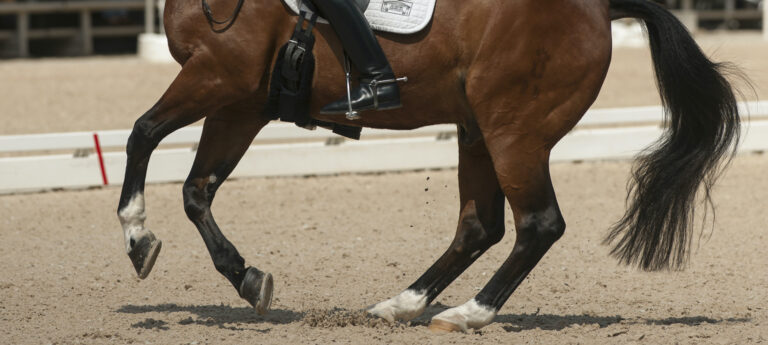
If a closer look at the respiratory system doesn’t reveal any obvious abnormalities, the next body system worth investigating is the musculoskeletal system. In many cases, what is perceived as fatigue is a sign of muscular disease, or myopathy. A typical case of tying up, called rhabdomyolysis, is easy to recognize. A horse suddenly becomes unwilling or unable to move, his muscles become hard and his gait very stiff and spastic. However, some myopathies are subclinical diseases, operating in the background to subtly undermine optimal performance. Because they don’t cause the classic overt signs of tying up, they go unrecognized. A simple step toward diagnosis is to measure the muscle enzyme levels in the horse’s blood. More detailed investigation involves taking a muscle biopsy.
There are a multitude of myopathies that have been categorized in the last decade, some with a genetic basis, and others of a less-characterized nature. The biopsy can clarify if there is a specific cause—for example, polysaccharide storage myopathy (PSSM), a genetic disorder that affects how a muscle stores and uses sugar. In some cases there is no name for the kind of myopathy, but examination of the muscle tissue confirms the chronic inflammation and can still help direct therapy.
Therapeutic strategies focus more on management. Common interventions include a more consistent exercise regimen, a carefully controlled dietary starch intake, switching to a ration with increased dietary fat and adding supplements to help muscle recovery after intense training. Additional interventions can be directed at replenishing fluid losses after training or before stressful events such as long-distance transport. Keep in mind that these are all general strategies, and the ultimate choice of management is dependent on the exact diagnosis.
Check His Heart
Another consideration for the source of exercise intolerance is the heart. Careful examination of the heart using a stethoscope in a quiet barn aisle is the first step to identifying a problem. This assessment can detect abnormal sounds called murmurs that are created by defects in the heart valves, as well as an irregular rhythm of the heartbeat, which is known as an arrhythmia. A thorough physical exam will assess the strength of the peripheral pulses in the feet or under the jawbone (the mandible). Venous distension (bulging blood vessels) or stocking up of the lower limbs and abdomen can be other signs that the heart isn’t functioning at its normal capacity.
If a cardiac problem is suspected, then an echocardiographic examination of the heart is indicated. Though this noninvasive ultrasound procedure can be performed in a horse’s home environment, many clients ship their horses to a referral clinic or university setting since these evaluations require an experienced practitioner and a powerful ultrasound machine to make a thorough assessment. The veterinarian places the ultrasound probe on the chest wall to visualize the heart between the ribs and evaluates the different chambers of the heart, its valves and outflow tracts, and can measure how well the heart contracts to expel the blood. These parameters can then be monitored over a horse’s lifetime to determine if the heart disease is progressing.
In cases of arrhythmias, additional analysis of the electrical signals coming from the heart over a longer period of time may be indicated. In this case, the horse is fit with a wearable device that collects heart rhythm data for 24 hours, and the horse can even be exercised on a high-speed treadmill while being recorded to know how the heart responds to intense training. Ultimately, the data is reviewed and a complete understanding of the relative risks of the abnormalities identified can be made. Many murmurs and even mild arrhythmias carry a reasonable prognosis for sport. Careful monitoring can help guide an owner to reduce a horse’s workload.
As you can see, identifying the cause for your horse’s recent lack of exercise tolerance may take a few steps, but there is usually a remediable solution. And the starting point is relatively straightforward: an examination by your veterinarian.
This column has not been approved or endorsed by U.S. Equestrian.
Christina “Cricket” Russillo, DVM, graduated from the Tufts University School of Veterinary Medicine in 2001. After completing a large animal medicine and surgery internship at Texas A&M, she realized her desire was to work on elite sporthorses. Following 13 years of practice at Fairfield Equine Associates in Newtown, Connecticut, focused on high-level show-jumping and dressage horses, she joined Virginia Equine Imaging in 2015. Russillo relocates to Florida every winter to support her clients and patients. She has competed through Third Level in dressage and in February 2017 she was appointed the U.S. Dressage Team veterinarian.