
Traditionally, when a horse’s gait feels off or lacks its usual impulsion, it is often assumed to be an issue of lameness. That, however, is not always the case. Thanks to improved diagnostic imaging tools and techniques now readily available to veterinarians, they are able to more accurately pinpoint problem areas that produce a decline in performance. Perhaps not so surprisingly, it’s not always in the legs or hooves that are the sites of the problem. With increasing frequency, the neck is being diagnosed as the root of common lameness, soreness, and performance issues.
The Anatomy of a Neck and What Can Go Wrong
In order to understand the problems that can arise in association with the horse’s neck, it’s important to first understand the anatomy.
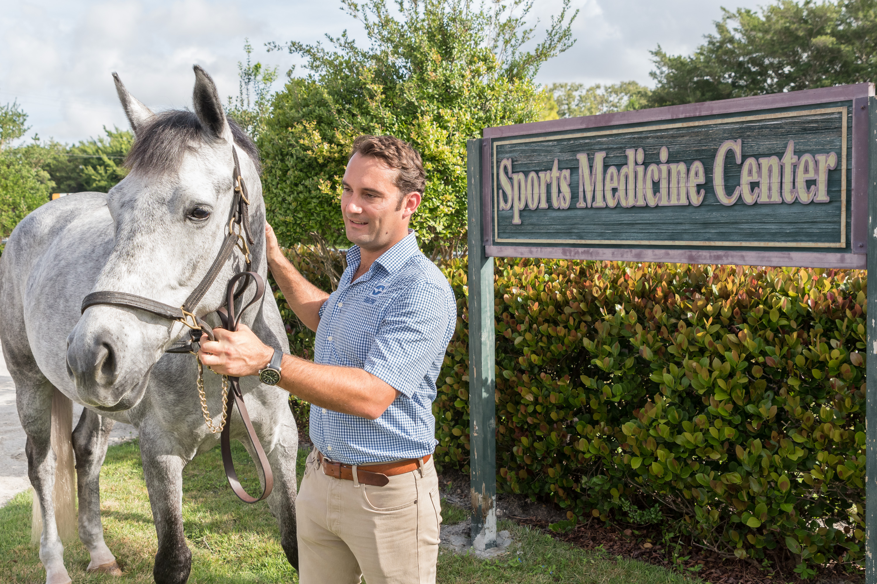
“The neck is composed of seven cervical vertebrae running from the head to the thorax, named C1 through C7, and each articulating with each other,” said Dr. Richard Wheeler, BVetMed, MRCVS, at Palm Beach Equine Clinic based in Wellington, FL. “The primary purposes of the neck are to move the head and to protect and transport the spinal cord and nerves, which run through the middle of the vertebrae.”
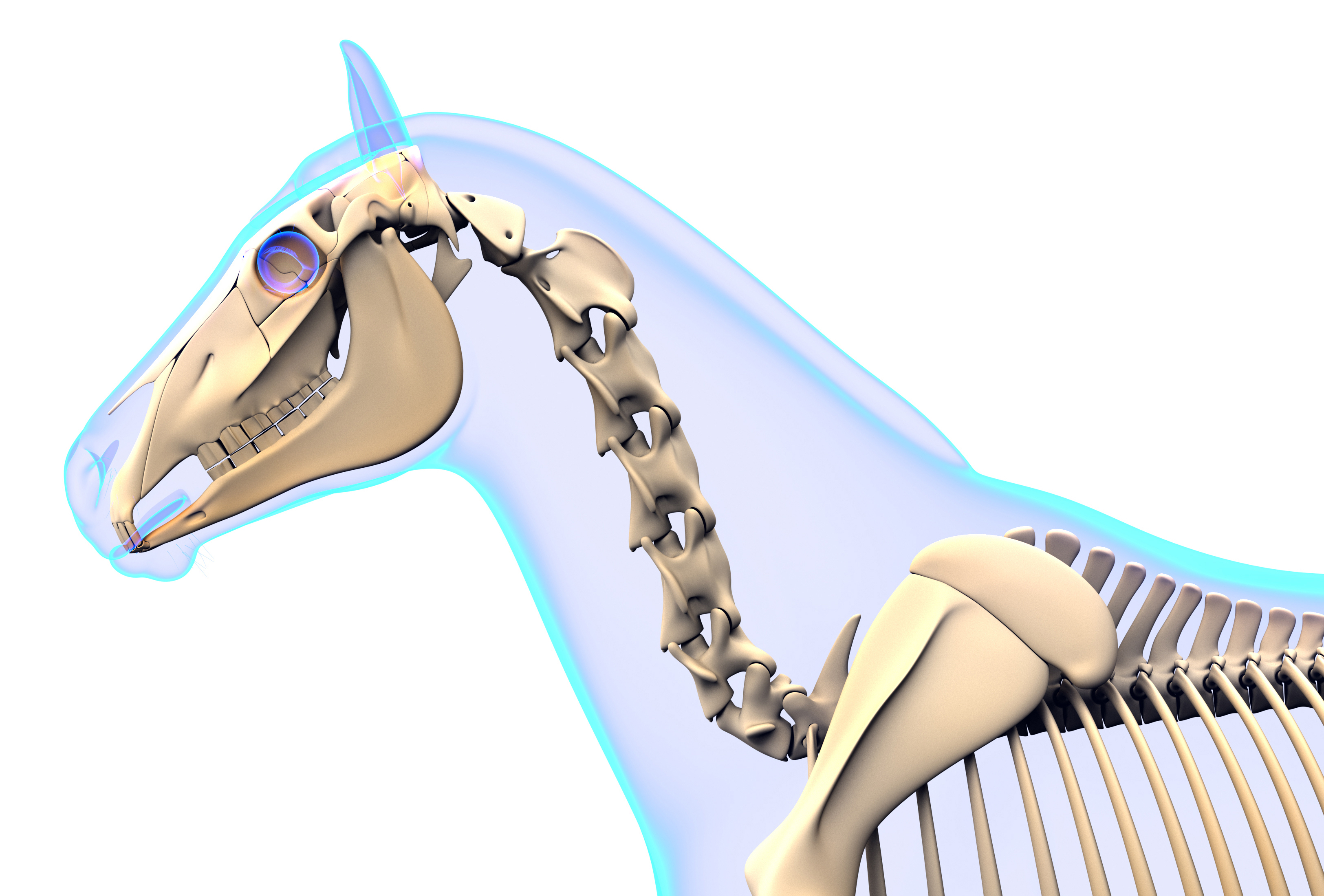
Such a major role as the protection of the nerves and spinal cord can also come with some major risks and complications, with clinical signs of these problems generally presenting themselves either neurologically, as neck pain, or as lameness in the front legs. These more specific symptoms may include:
Ataxia or clumsiness – Ataxia is defined as the “lack of control of bodily movements.” In the case of an ataxic horse, you may begin to notice staggering, sudden loss of balance, or even an inability to remain upright. Ataxia is generally an indicator of a neurological condition or damage to the spinal cord itself, caused by either developmental issues, trauma, or an infectious disease such as equine protozoal myeloencephalitis (EPM). Such neurological cases can often be the most debilitating.
Lameness – You can think of the spinal cord and the nerves in the neck like an interstate, with the spinal cord itself acting as the major highway. As you are “driving” along the interstate, every so often there are exits, which is where the nerves meet the cord. Should there be any impingement on the spinal cord itself, you’re likely looking at more severe complications – much like an accident on the highway. Should there be impingement on the nerves coming off of the spinal cord, it will more likely present itself like an accident a little way off an exit – not affecting the interstate itself, but possibly causing problems that spread elsewhere. That is when we see lameness issues arise.
Lameness resulting from the neck can difficult to pin down, but is often due to pressure on the smaller nerves that pass through the openings in the vertebrae and supply the front legs. Arthritis of the articular facet joints of the vertebrae is another common reason for lameness, because any time these joints become arthritic or inflamed, it can easily translate to the forelimbs.
Neck Pain – This will often go hand-in-hand with lameness, as factors such as arthritis of the articular facet joints can lead to both symptoms. Other possible reasons for neck pain include trauma or inflammatory diseases. While pain in the neck is not usually the first option considered when a horse turns up lame, but the pain can be the source of unexplained lameness and obvious discomfort.
Diagnosing the Problem
“The neck is one of the areas that has most clearly benefitted from the progression in advanced imaging,” said Dr. Wheeler. “While neck problems have likely been prevalent for some time, the veterinarians of Palm Beach Equine Clinic are now finding the diagnosis more common, as they are able to more accurately locate the issue.
“Neck problems, particularly those related to lameness, are generally diagnosed through a process of exclusion, nerve blocks are performed first to rule out lower regions of the horse’s body,” continued Dr. Wheeler. “Palpation of the neck, testing of the neck’s movement, and full neurological exams may also be performed in addition to a full lameness exam, depending on the horse’s symptoms.”
Once other regions of the horse are ruled out as the location of the problem, veterinarians are now able to use diagnostic images such as radiographs, nuclear scintigraphy, and standing computed tomography (CT) scans to specifically locate problems in the neck like never before.
“In years past, those diagnostic resources were left for last-ditch cases when veterinarians really could not pinpoint any other problems,” admitted Dr. Wheeler. “Today, with the advent of modern technology, better radiographs, better ultrasound machines, and more advanced diagnostic imaging from nuclear scintigraphy and CT scans, veterinarians are able to save time and money while finding the root of the problem more quickly and accurately.”
As an example, if arthritic factors are suspected, nuclear scintigraphy can be used to look for areas of increased bone turnover. On the resulting bone scan, veterinarians are looking for areas where there is an increased calcium uptake because the bone is actively remodeling. These areas will appear darker on the scan and are generally a good indicator of a boney injury or arthritis, with the darker “hot spots” often appearing above the articular facet joints.
In addition, groundbreaking advances for the diagnosis of equine neck problems include the use of a CT scan, with the ability to scan a standing horse with light sedation. A CT machine is one tool Dr. Wheeler’s arsenal of diagnostic imaging equipment at Palm Beach Equine Clinic.
Treatment
Once a solid diagnosis is arrived upon, the proper treatment protocols can be prescribed. Depending on the root of the problem, possible treatments may include shockwave therapy, regenerative therapies such as interleukin-1 receptor antagonist protein (IRAP) therapy, platelet-rich plasma (PRP) therapy, or one of the most common treatments, injections of the facet joints.
In the case of facet joint injections, veterinarians are able to medicate under ultrasound guidance, placing a needle into the joints and delivering corticosteroids or a similar medication. Surgery is also an option as a final approach to severe complications.
In milder cases, treatment may also all for increased time off, chiropractic treatments, or the administration of non-steroidal anti-inflammatory (NSAID) medications.
A problem within a horse’s neck is often underestimated as a source of lameness, decreased performance, and change in attitude or demeanor. Thanks, however, to the significant improvements in diagnostic and treatments technologies at clinics like Palm Beach Equine Clinic and others throughout the world, the equestrian community has another means to crack the code on undiagnosed problems. Just like in human medicine, veterinary medicine is evolving quickly and that progression is yielding more and more beneficial diagnostic options and effective treatments. These advances are a welcomed relief for horse owners and a saving grace for performance and companion horses alike.