Horses come in many shapes and sizes, and while all can benefit from basic dressage training, not all are physically capable of performing at the higher levels. One of the factors that can limit performance potential is conformation—the geometry of the skeletal framework in terms of the lengths and angulations of the bones and joints.
Ideally, the pelvis of a dressage horse should be long to give a large area for attachment of the propulsive muscles, and it should have a moderate slope to facilitate tilting the pelvis, lowering the haunches and moving the hind legs forward under the horse’s body.
Given the relationship between form (conformation) and function (performance), it is definitely helpful for the dressage rider to develop an eye for conformational features that affect a horse’s potential for dressage. The purpose of this article is to describe key features of the anatomy and conformation of the hindquarters with the goal of helping readers to evaluate important conformational features in this area.
Identifying Conformation
One of the secrets to being a good evaluator of conformation is to develop the skill to see beyond the outer layers of skin, fat and muscle in order to visualize the lengths and angles of the bones that lie beneath. Visualization of the bone structure is easier in some parts of the body than others. For example, below the elbows and stifles it is easy to see the outlines of the bones and to assess their conformation. In the shoulder and hip regions, however, the bones are concealed beneath the large, powerful muscles that attach the limbs to the trunk, making the bone structure more difficult to evaluate.
With that in mind, let us focus on the horse’s hindquarters and the differences between the shape of the croup versus the dimensions of the pelvis. The external contours of the croup are easy to see, but they give little information about the underlying pelvic structure, which is more relevant in our conformational evaluation.
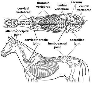
Let’s start by taking a look at the bones that underpin the structure of the hindquarters: the sacrum and the pelvis (see Figure 3 below). The sacrum is part of the vertebral column located between the lumbar region and the tail. Although there are five sacral vertebrae, they are fused together into a single bone, the sacrum, which means that there cannot be any movement between them. The joint between the front of the sacrum and the last lumbar vertebra—the lumbosacral joint—is quite mobile. Its movements can tilt the sacrum and pelvis forward (flexion or rounding), flatten the croup (extension or hollowing) or turn the haunches to the side (bending).
Figure 1 shows the anatomy of the sacrum from the side view. The underside is formed by the fused bodies of the five sacral vertebrae with their five spinous processes protruding upward. The spines on the sacrum get shorter from front to back. The tips of these spines form the topline in the middle of the horse’s croup.
The orientation (slope) of the sacrum varies with the horse’s posture. Horses with good strength and tone in their core musculature hold the lumbosacral joint in a slightly flexed position and keep the croup slightly tucked. Loss of tone in the core musculature may be reflected in poor posture even to the extent that the pelvis slopes
upward toward the tail head.
As you can see in Figure 1, the amount of shortening of the sacral spines varies between horses, and this also affects how much the croup slopes downward toward the tail head. Note that in the live horse, the gluteal muscles may bulge on either side of the sacral spines giving a rounded profile, so it is important to assess the slope of the croup (sacrum) on the midline between the gluteal muscles.
Immediately behind the sacrum are the vertebrae of the tail. The length and angulation of the sacrum affect the position and carriage of the tail. A flat (horizontal) croup is associated with a high tail set and a high tail carriage as shown by the Arabian in the top photo in Figure 5. A sloping croup gives a lower tail set and a lower tail carriage as shown by the Friesian in the top photo in Figure 6.
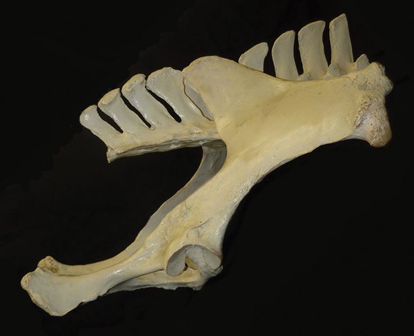
The pelvis connects the bones of the hind limb to the vertebral column via the hip joint and the sacroiliac joint. When seen from behind, the pelvis is somewhat U-shaped with a narrow separation in front where it curves around and above the sacrum (see Figure 2). The union between pelvis and sacrum at the left and right sacroiliac joints suspends the sacrum beneath the pelvis and anchors it in place with strong ligaments. The sacroiliac joints, which are located on either side between the high points of the croup, do not allow a significant amount of movement; their function is to transmit propulsive forces generated by the hind limbs.
The hip joints are on either side of the lower part of the pelvis, where the acetabulum forms a rounded socket that receives the head of the femur. The hip is a highly mobile joint that allows the entire hind limb to swing back and forth and to move sideways in abduction (swinging outward) and adduction (swinging inward). A low-set hip joint facilitates compression of the hip angle and is advantageous for allowing the horse to perform highly collected movements.
Pelvic Length & Angulation
The pelvis is surrounded by the large muscles of the hindquarters, making it difficult to distinguish the contours. But, fortunately, there are three bony prominences on each side that are easy to see and feel and that we can use as landmarks to assess pelvic conformation. These are the point of the hip (tuber coxae), the point of the buttock (tuber ischii) and the point of the croup (tuber sacrale). Pelvic length and slope are measured by drawing a line from the upper part of the point of the hip to the point of the buttock, which is a few inches below the tail head (Figures 2 and 3).
Both the length and angulation of the pelvis are key conformational measurements that affect the horse’s strength, power, speed and agility. A larger (longer and broader) pelvis has more room for attachment of the powerful gluteal and hamstring muscles that provide propulsion during locomotion.
Horses that race over short to middle distances, such as racing Quarter Horses and Thoroughbreds, have the longest pelvises, measuring up to one third of the total body length. A short pelvis offers less area for attachment of the propulsive muscles, but this is compensated by greater agility. Dressage horses have pelvises that tend toward the longer end of the spectrum, though not as long as racehorses.
The angle of the pelvis is measured relative to the horizontal with the horse standing square. Using these landmarks, an average angle for a dressage horse’s pelvis would be around 20 degrees. In his doctoral research, Swedish equine biomechanics expert Dr. Mikael Holmström found that the average pelvic angle in elite Swedish Warmblood dressage horses was 30 degrees. However, it should be noted that Dr. Holmström measured pelvic angle from the upper part of the point of hip to the hip joint.
These landmarks will always give a steeper pelvic angle than if it had been measured from the point of the hip to the point of the buttock. It’s not a matter of one method being right or wrong; it’s just two slightly different measurement techniques. However, you need to know which landmarks were used in order to evaluate and compare the results.
When the lumbosacral joint is flexed, the rear part of the pelvis tilts forward, bringing the hip joint and hind leg farther forward under the horse’s body. Equine conformation analysis expert Dr. Deb Bennett refers to this as “coiling the loins,” which helps us to visualize the effect. With the pelvis tilted forward, the frame is compressed and the hind limbs act closer to the center of gravity, providing more upward (rather than forward) propulsion. A significant amount of pelvic tilting (and untilting) occurs during each stride of canter when the lumbosacral joint flexes as the hind limbs are pulled forward and extends as they are retracted.
In piaffe and canter pirouettes, the horse can maintain lumbosacral flexion and keep the pelvis tilted forward throughout the stride because in these movements the hind limbs are not retracted. In horses with a flat pelvic conformation, the sublumbar muscles that are responsible for lumbosacral flexion exert less leverage than in horses with a more sloping pelvic conformation. Thus, it requires greater force to flex the lumbosacral joint with a flatter pelvic angle, and lumbosacral flexion compresses the hip downward, rather than tilting it forward. On the other hand, an overly steep pelvis may restrict the rearward swing of the hind limb and interfere with the ability to extend the stride. As with many conformational variables, extreme pelvic angulations in either direction are not ideal; and an intermediate angle is preferred.
In recent years, selective breeding for specific performance criteria has produced horses that excel in dressage, though these superstars are out of the reach of most riders. It is possible, however, to find horses of a variety of breeds that have conformation favorable for dressage. The key is to learn how to distinguish between horses that have the physical attributes needed for dressage versus those that are better suited to another occupation.
The breed photographs in this article compare the conformation of the croup and pelvis of an outstanding warmblood dressage competitor (Figure 4) with two representatives each of the Arabian and Friesian breeds to show the diversity of croup conformation within these breeds (Figures 5 and 6). The photos show that there are horses in each of these nonwarmblood breeds that have suitable conformation to become good dressage performers.
Dr. Hilary Clayton holds the Mary Anne McPhail Chair at Michigan State University’s College of Veterinary Medicine in East Lansing. The goal of the chair is to perform scientific investigations that directly benefit the sport of dressage. A native of England, Dr. Clayton received her veterinary degree from the University of Glasgow in 1973. In 1982, she accepted a position with the University of Saskatchewan in Canada, where she spent 15 years as a professor of veterinary anatomy. As a veterinarian and researcher, her studies on the biomechanics of equine gait have focused on sport horses. She is a U.S. Dressage Federation bronze, silver and gold medalist and is a certified equestrian coach in the United Kingdom and Canada. She has been a member of the Canadian National Coaching Committee for the sports of dressage, jumping and eventing, and is currently a member of the U.S. Equestrian Federation’s Dressage Committee.

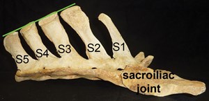
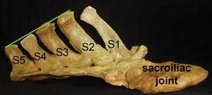
Figure 1: Difference in Shape of the Sacrum
The bones are positioned so that the head of the horse would be to the right and the tail to the left. The sacral spines are labeled S1 to S5 and the green line shows the croup angle. These specimens illustrate how the size and shape of the bones vary between horses. In the top sacrum, the first sacral spine (S1) is short and poorly developed and the croup angle is 24 degrees. In the bottom sacrum, there is a more marked difference in length between the second (S2) and last (S5) spines and the individual spines have more of a backward slope. The croup angle is 30 degrees. These are examples of the diversity seen among normal horses.
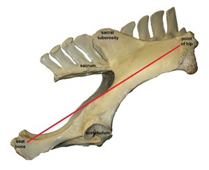
Figure 2: Horse Pelvis Articulated with the Lumbar Vertebrae and Sacrum.
The horse’s head would be to the right and the tail to the left. The three bony prominences have been labeled: point of croup, point of hip and point of buttock (see Figure 3). The acetabulum is also labeled; it forms the articulation of the hip joint. A lower position of the acetabulum favors the ability to perform highly collected movements.
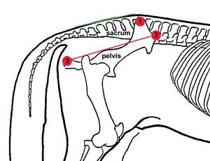
Figure 3: The Position of the Sacrum and Pelvis Relative to the Contours of the Hindquarters
The three prominences on the pelvis are marked by red circles: 1. point of croup; 2. point of hip; 3. point of buttock. The red line running from the point of hip to point of buttock indicates the slope of the pelvis. The green line indicates the slope of the croup. In this diagram the two slopes are approximately the same. This horse has a rather flat (horizontal) croup and pelvic angles, and the acetabulum is placed relatively high.
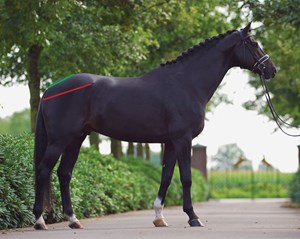
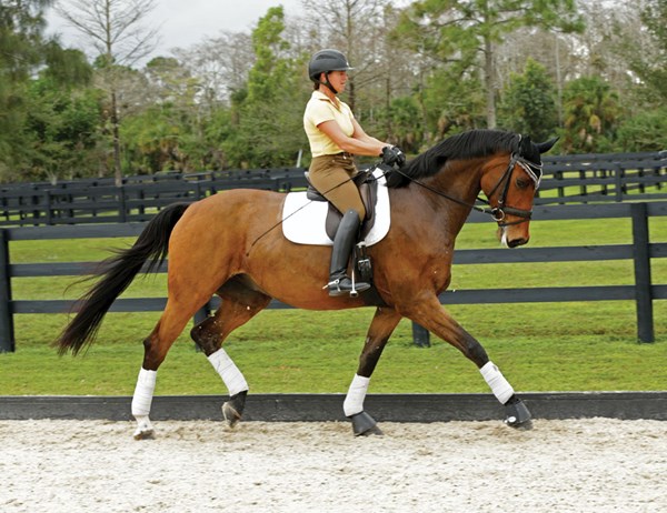
Figure 4: The Warmblood
The warmblood is often considered the standard for ideal dressage conformation. The photo below illustrates how the hindquarters of successful dressage horses often have pelvises that tend toward the longer end of the spectrum and have a moderate slope.
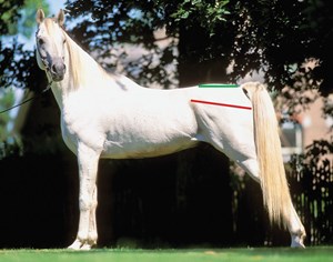
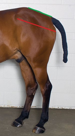
Figure 5: The Arabian
These two Arabians are of very different types. The top horse is a successful halter horse and is posed with the hind limbs camped out and the lumbosacral joint extended to make the croup appear as flat as possible. In this horse the croup angle is horizontal and the pelvic angle is 10 degrees. The high tail set and high tail carriage are also exaggerated in this pose. If this horse were not standing camped out, both the croup and pelvic angles would be a little more sloped. Neither the excessively flat croup nor the tendency to stand and move with the lumbosacral joint extended are desirable characteristics in a dressage horse. The lower photo is of a successful Arabian Grand Prix competitor. The angles of the croup and pelvis are parallel and measure 20 degrees. Perhaps unusually for an Arabian, this horse has a good ability to tilt the pelvis forward and engage the hind limb.
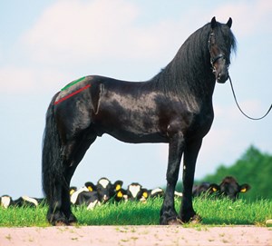
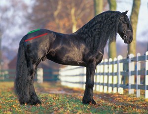
Figure 6: The Friesian
Friesians were originally bred for use in warfare and agriculture. The breed is still popular as a light carriage horse, and the top Friesian photo is an example of the Baroque type used for driving. Note the steeply sloped croup and pelvis (both 20 degrees) and the consequent low-set tail.
The Friesian sport-horse bloodlines, as illustrated by the horse in the bottom photo, are becoming increasingly popular for dressage. This Friesian sport horse has greater length and less slope in the hindquarters compared with the horse above.


